Author: Jaqueline Tran, MD
Peer Editors: Lauren Gruffi, MD and Amy Hembree, MD
“B-Teal Tachycardia coming back”
Introduction
Atrial fibrillation with rapid ventricular response (AF RVR) is a disease entity that we as emergency physicians encounter daily. AF itself comprises 0.5% of all ED visits and remains the most common dysrhythmia managed in the ED. [1]
AF RVR is associated with significant morbidity and mortality, often associated with high rates of hospital admission. The typical patient presenting to the ED with acute or rapid Afib is typically older (mean age of 69-70), male, white, and with multiple comorbidities including hypertension diabetes, vascular disease, and congestive heart failure. [2]
In medical school, afib management was made easy: Unstable? Shock. Stable? Rate control. While this barebones approach was good enough for the occasional Step 2 question, it is inadequate–and quite frankly dangerous–in a real life ED context. In this post, we are going to explore the various ways in which AF RVR is deceptively difficult and nuanced to manage.
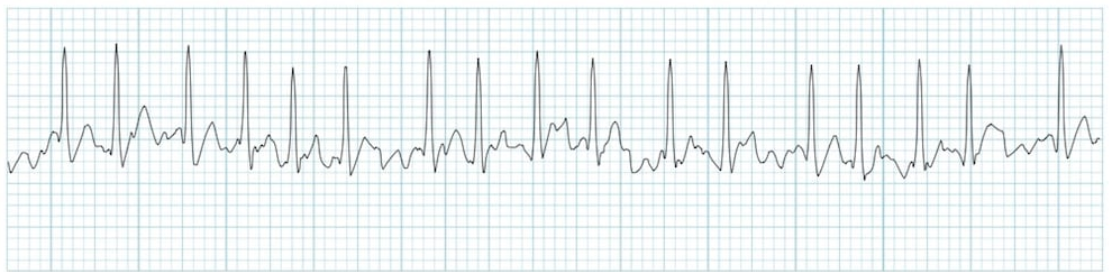
Figure 1. Irregularly irregular EKG pattern of atrial fibrillation
Pathophysiology
Atrial fibrillation arises from disorganized atrial electrical activation (most commonly originating from the pulmonary veins) without coordinated atrial contraction, accompanied by irregular ventricular response. As a result, it produces an EKG pattern described as irregularly irregular, as shown above in Figure 1. Over time, persistent and long-standing AF triggers structural remodeling of the atria as well as functional remodeling of the sinus and AV node, which further perpetuates AF (Figure 2). Consequently, patients with AF suffer from left atrial dilation, left ventricular dysfunction, and decreased cardiac output. While there is no universal heart rate threshold to define “rapid,” the term “RVR” most often comes into play once the heart rate is >110. [3]
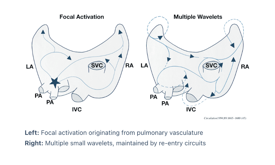
Figure 2. Electrophysiological concepts demonstrating how afib begins and perpetuates.
Management:
There are 5 simple questions I ask myself every time I have a patient in AF RVR:
Are they stable?
Why are they in AF RVR?
Do I want to give fluids?
What do I think about rate control?
What do I think about rhythm control?
1. Are they stable?
What we really mean by this is: what is the patient’s blood pressure and mental status? How rapid is their rate, and how well are they compensating? An asymptomatic AF with a rate of 115 is significantly less concerning to me than symptomatic AF with a rate of 186. Blood pressure is not the only marker we use to determine inadequate perfusion, and a blood pressure of 90/50, while technically “hypotensive” does not always make me immediately reach for the pads–which leads me to the next point.
2. Why are they in AF RVR?
While AF RVR can present as a primary disease, it is very often a secondary, compensatory process. If a patient’s AF RVR is a consequence of an underlying pathology, treatment of the driving force can often terminate the arrhythmia. Jumping to control the rate or rhythm with electricity or pharmacologic agents becomes a bandaid–and when the AF RVR may actually be helping a patient compensate, artificially controlling the rate or rhythm can have dire consequences for their hemodynamics. For example, the floridly septic patient with a HR of 165 and BP 80/40 is likely in a distributive shock and needs to be fluid resuscitated, NOT to be cardioverted (despite them being in “unstable AF RVR”!) Some other common triggers for AF RVR in the ED include electrolyte imbalance, alcohol use, hyperthyroidism, acute MI, and pulmonary embolism.
3. Do I want to give fluids?
How do I know? POCUS! The ultrasound is, in my opinion, the single most useful diagnostic tool at your disposal in the acute setting. Within minutes, an ED provider can perform 1) focused cardiac and pulmonary POCUS (evaluate for EF, pericardial effusion, signs of right heart strain, pulmonary edema) 2) evaluate the IVC for fluid responsiveness 3) FAST or RUSH exam. Knowing any of this information will determine your next steps in management.
4. What do I think about rate control?
Should I choose rate control? Beta blockers vs Calcium channel blockers? IV or PO? Will cardiology hate us? All great questions. Knowing when to implement rate control in AF RVR can be tricky.
Contraindications:
According to the American College of Cardiology (2024), intravenous nondihydropyridine calcium channel blockers should not be administered to patients in AF RVR with known, reduced LV systolic function (<40%) or decompensated heart failure (Class III: Harm recommendation) due to their negative inotropic and chronotropic effects.
Nondihydropyridine calcium channel blockers (such as diltiazem) exert negative inotropic and chronotropic effects by inhibiting calcium influx through voltage-gated L-type calcium channels, thereby inhibiting sarcoplasmic reticulum release of intracellular calcium. This reduces available calcium substrate for excitation-contraction coupling (inotropic) and conduction tissue depolarization (chronotropy) in cardiac muscle cells. It is worth noting that dihydropyridine calcium channel blockers, such as amlodipine and nifedipine, are not considered in rate/rhythm control for tachydysrhythmias due to their specificity for arteriolar, rather than cardiac, tissue.
Similarly, beta blockers are contraindicated in decompensated heart failure. By blocking B1-adrenergic receptors in the myocardium, beta blockers have negative inotropic effects that reduce myocardial contractility, and it is this sympathetic drive and subsequent tachycardia (or AF RVR) that is likely helping to maintain the patient’s cardiac output.
An additional consideration with beta blocker usage is the presence of reactive airway disease, as initiating beta blockade may worsen bronchospasm in susceptible patients. Agents such a diltiazem are therefore often the agent of choice in patients with a history of asthma or COPD and no history of heart failure.
Importantly, if a patient has a known preexcitation syndrome (ie WPW), be wary of AV nodal blocking agents as it may precipitate ventricular fibrillation (vfib).
Medication Choice
You’ve now determined that your patient is an appropriate candidate for rate control -- what are you reaching for? The reality is that the age-old beta blocker vs calcium channel blocker debate is still not settled. A 2022 meta-analysis of 17 studies suggest IV diltiazem to have some advantages over IV metoprolol in achieving rate control (shorter onset time by 1.13 minutes, lower achieved ventricular rate by 9.48 BPM) without significant difference in adverse events. Yet, a 2025 systematic review and meta-analysis specifically investigating adverse events–defined as hypotension or bradycardia–found that metoprolol was associated with 26% lower risk of adverse events (hypotension or bradycardia) (total incidence 10%) compared with diltiazem (total incidence 19%) (RR 0.74; 95% CI 0.56–0.98, p=0.034). [5] Importantly, there has been no identified mortality benefit with either beta blockers or calcium channel blockers. Thus, medication selection typically weighs the amount of time needed to achieve rate control (diltiazem > metoprolol) against the potential for hypotension and bradycardia (metoprolol > diltiazem). (If you’ve dealt with AF RVR in the ED enough times, you will surely have experienced the “we tried metoprolol 3 times and didn’t work, but then dilt worked like magic! Andddd their blood pressure also tanked” scenario). This is because beta blockers and calcium channel blockers have additive effects on the heart; they both slow conduction through the AV node, decrease myocardial contractility, and decrease cardiac output.
Metoprolol is the most commonly utilized beta blocker due to its availability in IV and PO formulations, provider familiarity, and cardioselectivity. Agents like propranolol are nonselective, and agents like esmolol (while effective) are IV only and administered as continuous infusions requiring constant monitoring. The standard initial dosing of metoprolol is 2.5-5mg IV bolus over 2 minutes, up to 3 times. The half life is approximately 3-4 hours, thus it is prudent to follow the IV bolus with PO dosing to prevent recurrence of tachycardia. The AHA recommends initiation of PO therapy 15 minutes after the last IV bolus at 25-50mg q6h for the next 48h before being transitioned to maintenance dosing.
Diltiazem is the most commonly utilized calcium channel blocker due to its availability in both IV and PO formulations and provider familiarity. The AHA has noted that while verapamil is an option, there is a lack of clinical trials studying the use of verapamil in acute settings. Standard initial dosing of diltiazem is 0.25mg/kg IV bolus–if rate control is not achieved after 15 minutes, it can be followed by a 0.35mg/kg IV bolus. Like metoprolol, IV diltiazem can be followed by 30-60mg immediate release PO diltiazem. Previous management encouraged initiation of a continuous diltiazem infusion for rate control maintenance; however, a recent 2025 study published in AJEM noted no significant difference in treatment failure at 4 hours when comparing PO diltiazem with continuous diltiazem infusion, and a higher rate of ED discharge when patients were initiated on PO (31.7% vs 4%). [4]
Don’t sleep on magnesium! A 2021 meta analysis comparing standard rate control therapy (beta blocker and calcium channel blocker) vs standard + IV magnesium was superior in achieving rate control (63% to 40%, OR 2.49, CI 1.8-3.45) and with rhythm conversion (21% vs 14$, OR 1.75, CI 1.08-2.84). With little side effect and risk of harm, magnesium is an excellent adjunctive therapy. [6]
5. Rhythm Control
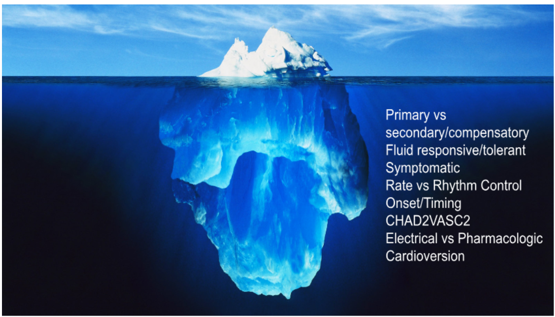
Amio? To shock or not to shock? When? This decision, like all others mentioned above, is nuanced. “Hypotensive AF RVR = shock” is not always an appropriate response, and all of the considerations (primary vs compensatory AF RVR, fluid status, CHF status) that we discussed above continue to play a role.
Rhythm control is an appropriate consideration when it is thought that the AF RVR is the driving factor in a patient’s hemodynamic instability, and when rate control is inappropriate or contraindicated, in a patient.
A number of factors come into play when considering rhythm control, including:
Duration/timing of onset of AF
Anticoagulation status
New onset AF vs known chronic AF
CHADVASC score
Ability to tolerate the intervention (ie sedation for electrical cardioversion)
Generally speaking, there is no hard and fast rule for the timing of cardioversion—various studies have cited the onset of AF less than 12 hours, 36 hours, or even 48 hours as safe to cardiovert when anticoagulation status is unknown. [7] If time of onset is truly unknown but cardioversion is desired,TEE remains the gold standard for evaluating the presence or absence of left atrial thrombi. However, Cardiac CT/CT Pulmonary Vein (done inpatient) can be a reliable alternative when TEE is not plausible. If TEE is not immediately available, consider concurrent initiation of IV heparin bolus and drip, for both electrical and chemical cardioversion.
While not directly used or validated for determining risk of thromboembolism (TE) in cardioversion, CHAD2VASC2 score can be calculated to help understand a patient’s background risk for TE in 1 year. [Figure 3]. For example, a male patient with a score of 0, or female patient with a score of 1, would be considered low-risk.
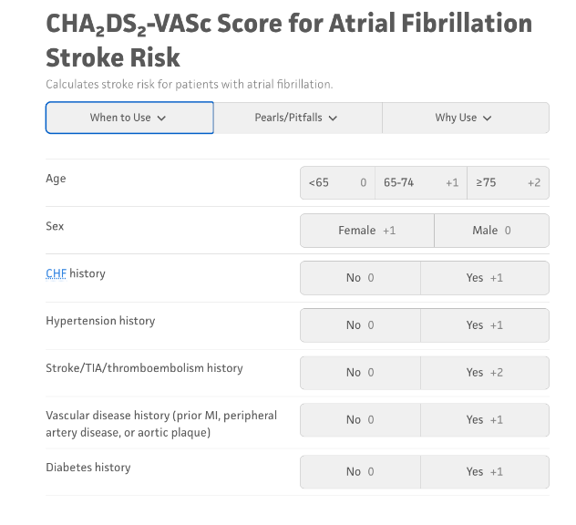
Figure 3. CHAD2VASC2 score for AF Stroke Risk
Pharmacologic vs Electrical Cardioversion
It is thought that electrical cardioversion has a higher success rate than pharmacologic cardioversion. However, it should be noted that studies have been limited by the lack of data in patients who are hemodynamically unstable. In retrospective studies on stable patients, DCCV has been reported to have a 78.2% success rate compared to pharmacological cardioversion (59.2%) according to the ACC. That being said, both pharmacologic and electrical cardioversion are considered reasonable options for rhythm control. [8]
Electrical cardioversion is ideally performed with the patient sedated, synchronized with the QRS complex and delivered at 200 joules. Pre-treatment with antiarrhythmics can help increase the chance of successful cardioversion. The benefits of electrical cardioversion are theoretically higher rate of success and the immediacy of rhythm conversion, which are ideal in unstable situations. However, the need for sedation, as well as patient apprehension with the procedure, are potential limitations to its use.
Amiodarone has been extensively studied and found to be effective in cardioversion, and does not require sedation unlike electrical cardioversion. It is given as a loading dose of 5-7mg/kg (max 300mg) followed by an infusion of 10-50mg/h for the next 24h. Downsides include onset of action, as it may take 8-12 hours for rhythm control. Important considerations when opting for amiodarone include liver metabolism, thyrotoxicosis, AV nodal blockade, and QT prolongation.
Key Takeaways:
AF RVR is not that simple! Look for a cause, treat it first if able. POCUS can answer most of your clinical questions in a few minutes.
Beta blockers and calcium channel blockers are contraindicated in decompensated HFrEF
Amiodarone works but can be slow, and it has a significant side effect profile
Electrical cardioversion works but patients are often apprehensive, and it requires sedation
Sometimes you learn through trial and error when you don’t have all the information, and that’s okay.
References:
Atzema CL, Austin PC, Miller E, Chong AS, Yun L, Dorian P. A population-based description of atrial fibrillation in the emergency department, 2002 to 2010. Ann Emerg Med. 2013;62(6):570-577.e7. doi:10.1016/j.annemergmed.2013.05.019.
Deshmukh A, Pant S, Kumar G, et al. Emergency department visits for atrial fibrillation in the United States: Trends in admission rates and economic burden from 2007 to 2014. J Am Heart Assoc. 2018;7:e009024. doi:10.1161/JAHA.118.009024.
Jeong JH, Baek YS, Park J, Park HW, Choi EK, Park JK, Kang KW, Kim J, Lee YS, Kim JB, Choi JI, Joung B, Shim J. Impact of Atrial Fibrillation with Rapid Ventricular Response on Atrial Fibrillation Recurrence: From the CODE-AF Registry. J Clin Med. 2024 Sep 14;13(18):5469. doi: 10.3390/jcm13185469. PMID: 39336961; PMCID: PMC11432096.
Agee JK, Baize RC, Nguyen M, et al. Evaluation of diltiazem dosing strategies in the management of atrial fibrillation with rapid ventricular response. Am J Emerg Med. 2025;98:154-160. doi:10.1016/j.ajem.2024.10.026
Hintze TD, Downing JV, Acquisto NM, Downton Mslis K, Yardi I, Moran M, Sharma A, Pourmand A, Tran QK. Metoprolol vs diltiazem for atrial fibrillation with rapid ventricular rate: Systematic review and meta-analysis of adverse events. Am J Emerg Med. 2025 Mar;89:230-240. doi: 10.1016/j.ajem.2024.12.070. Epub 2024 Dec 27. PMID: 39764905.
Ramesh T, Lee PYK, Mitta M, Allencherril J. Intravenous magnesium in the management of rapid atrial fibrillation: A systematic review and meta-analysis. J Cardiol. 2021;78(5):375-381. doi:10.1016/j.jjcc.2021.06.001.
Capranzano P, Calvi V. Timing of cardioversion in atrial fibrillation: the sooner the better? Eur Heart J Suppl. 2020 Nov 18;22(Suppl L):L41-L43. doi: 10.1093/eurheartj/suaa132. PMID: 33654465; PMCID: PMC7904082.
Writing Committee Members; Joglar JA, Chung MK, Armbruster AL, Benjamin EJ, Chyou JY, Cronin EM, Deswal A, Eckhardt LL, Goldberger ZD, Gopinathannair R, Gorenek B, Hess PL, Hlatky M, Hogan G, Ibeh C, Indik JH, Kido K, Kusumoto F, Link MS, Linta KT, Marcus GM, McCarthy PM, Patel N, Patton KK, Perez MV, Piccini JP, Russo AM, Sanders P, Streur MM, Thomas KL, Times S, Tisdale JE, Valente AM, Van Wagoner DR. 2023 ACC/AHA/ACCP/HRS Guideline for the Diagnosis and Management of Atrial Fibrillation: A Report of the American College of Cardiology/American Heart Association Joint Committee on Clinical Practice Guidelines. J Am Coll Cardiol. 2024 Jan 2;83(1):109-279. doi: 10.1016/j.jacc.2023.08.017. Epub 2023 Nov 30. Erratum in: J Am Coll Cardiol. 2024 Mar 5;83(9):959. doi: 10.1016/j.jacc.2024.01.020. Erratum in: J Am Coll Cardiol. 2024 Jun 25;83(25):2714. doi: 10.1016/j.jacc.2024.05.033. PMID: 38043043; PMCID: PMC11104284.